
23 LET’S PLAY THE BRAIN GAME
ALEX NORTON︎︎︎
Process
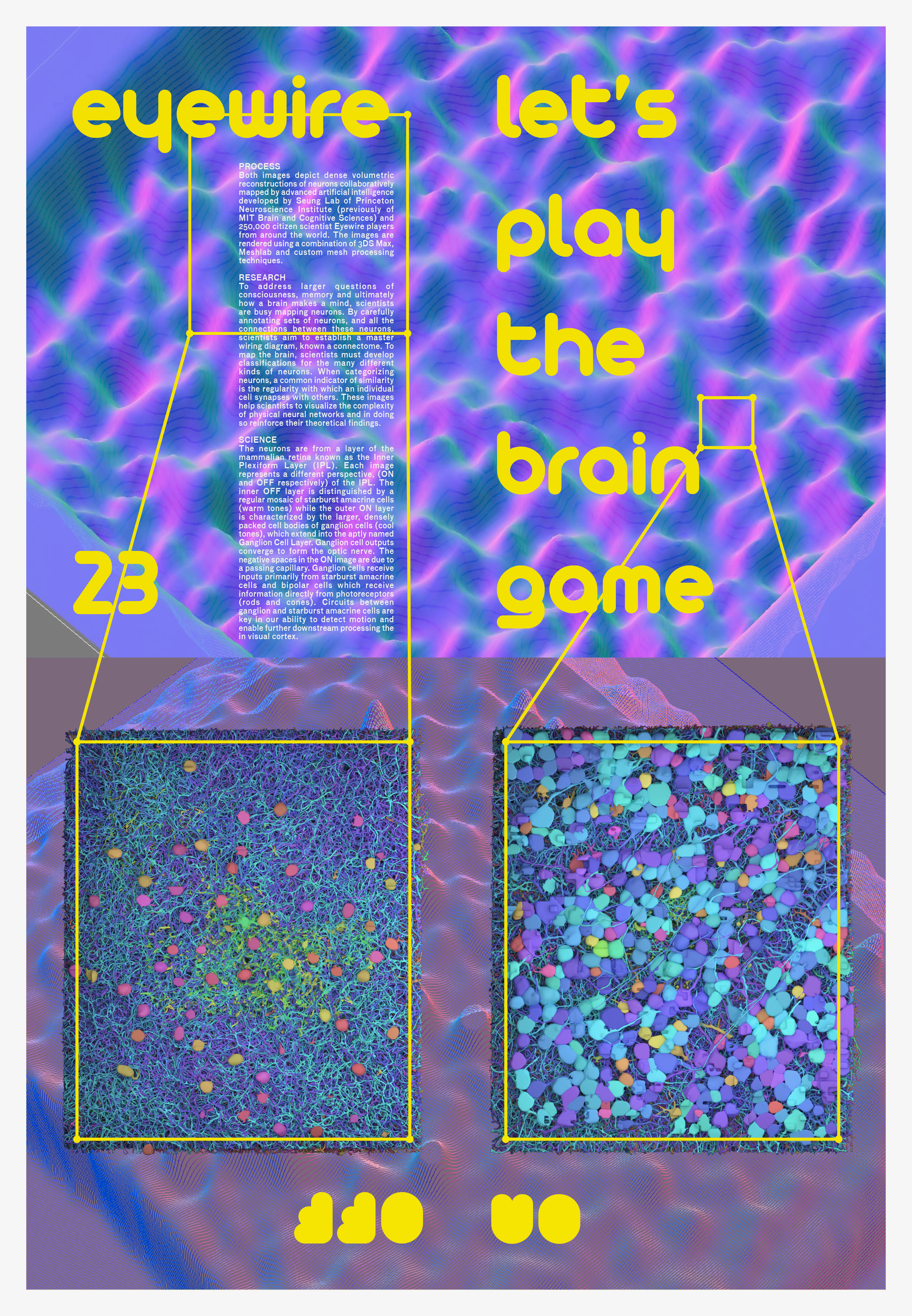
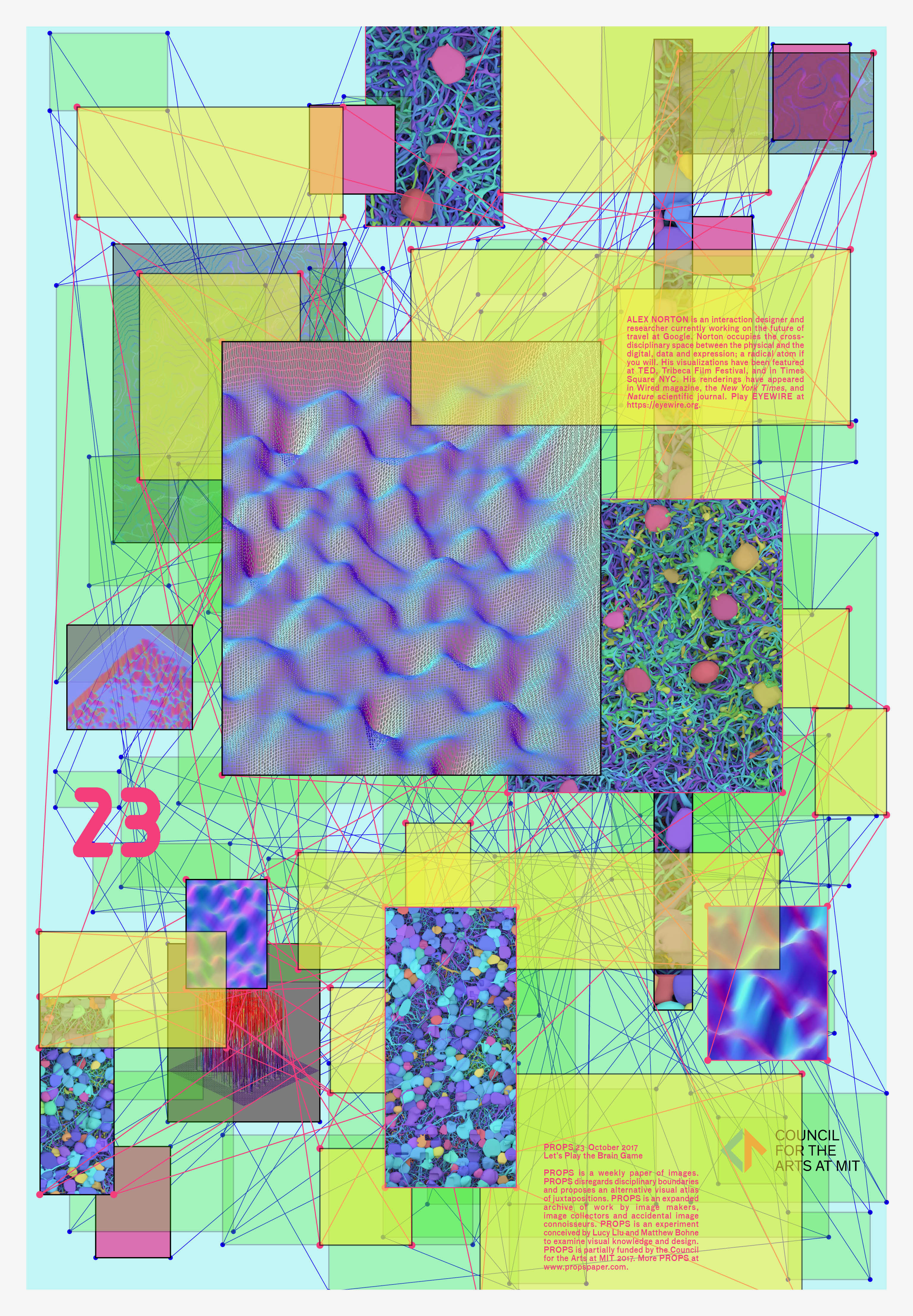
Both images depict dense volumetric reconstructions of neurons collaboratively mapped by advanced artificial intelligence developed by Seung Lab of Princeton Neuroscience Institute (previously of MIT Brain and Cognitive Sciences) and 250,000 citizen scientist Eyewire players from around the world. The images are rendered using a combination of 3DS Max, Meshlab and custom mesh processing techniques.
Research
To address larger questions of consciousness, memory and ultimately how a brain makes a mind, scientists are busy mapping neurons. By carefully annotating sets of neurons, and all the connections between these neurons, scientists aim to establish a master wiring diagram, known a connectome. To map the brain, scientists must develop classifications for the many different kinds of neurons. When categorizing neurons, a common indicator of similarity is the regularity with which an individual cell synapses with others. These images help scientists to visualize the complexity of physical neural networks and in doing so reinforce their theoretical findings.
Science
The neurons are from a layer of the mammalian retina known as the Inner Plexiform Layer (IPL). Each image represents a different perspective, (ON and OFF respectively) of the IPL. The inner OFF layer is distinguished by a regular mosaic of starburst amacrine cells (warm tones) while the outer ON layer is characterized by the larger, densely packed cell bodies of ganglion cells (cool tones), which extend into the aptly named Ganglion Cell Layer. Ganglion cell outputs converge to form the optic nerve. The negative spaces in the ON image are due to a passing capillary. Ganglion cells receive inputs primarily from starburst amacrine cells and bipolar cells which receive information directly from photoreceptors (rods and cones). Circuits between ganglion and starburst amacrine cells are key in our ability to detect motion and enable further downstream processing the in visual cortex.
ALEX NORTON︎︎︎ is an interaction designer and researcher currently working on the future of travel at Google. Alex’s visualizations have been featured at TED and in Times Square NYC. His renderings have appeared in Wired magazine, the New York Times, and Nature scientific journal. His work focuses on the intersection of people and technology, weaving lessons from natural systems into creative solutions, delightful interactions, and computational models. By harnessing the power of advanced 3D graphics, Alex communicates the impact of complex scientific findings to the public.